
Diabetes is a chronic illness that can cause a variety of complications, one of which is diabetic retinopathy. This eye condition arises when diabetes leads to damage in the blood vessels of the retina, the light-sensitive tissue at the back of the eye. If left untreated, diabetic retinopathy can progress to severe vision loss or even complete blindness. It is therefore imperative for individuals with diabetes to undergo regular eye examinations to monitor for early signs of this potentially sight-threatening disease.
Fundus photography is a crucial diagnostic tool used in the early detection of diabetic retinopathy. This method involves capturing detailed images of the back of the eye, specifically focusing on the retina. The resulting images allow eye care professionals to observe any changes or abnormalities that may signify the onset of diabetic retinopathy.
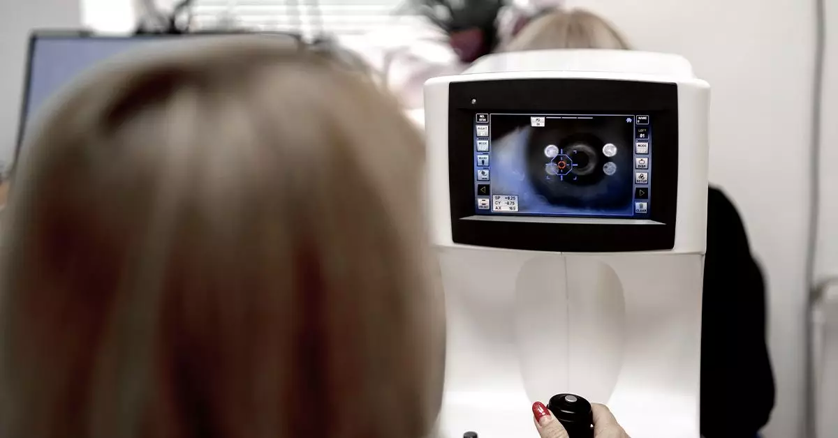
During a fundus photography session, eye care specialists typically use a specialized camera designed to capture high-resolution images of the retina. The procedure starts with the application of dilating eye drops to enlarge the pupils, facilitating a clearer view of the eye’s interior. The dilation process can take up to half an hour, and during this time, patients may experience temporary blurred vision or sensitivity to light.
What’s remarkable is that recent technological advancements have made it possible for patients to capture their own fundus images using smartphones. This innovative approach, known as Selfie Fundus Imaging (SFI), allows individuals to take pictures of their retinas after dilating their eyes at home. Research has shown that images taken via SFI can be as effective as those captured by professional equipment, significantly enhancing accessibility and affordability for diabetic retinopathy screening.
Several crucial signs of diabetic retinopathy can be detected through fundus photography, making the process invaluable for timely intervention. Some of the common indicators include:
1. **Microaneurysms**: These small, round, red spots appear on the retina, representing the earliest detectable lesions related to diabetic changes in the eye.
2. **Retinal Hemorrhages**: Damaged blood vessels can lead to bleeding within the retina, manifesting as flame-shaped or dot-and-blot hemorrhages, which are vital for diagnosis.
3. **Hard Exudates**: Formed by lipid deposits that leak from blood vessels, these yellowish, waxy patches can also serve as a warning signal.
4. **Cotton Wool Spots**: These fluffy white patches point to areas of the retina that are experiencing insufficient blood supply, indicating a growing concern.
5. **Intraretinal Microvascular Abnormalities (IRMA)**: These represent abnormalities within the retinal blood vessels, hinting at more serious vascular issues.
6. **Neovascularization**: Seen in advanced stages of diabetic retinopathy, this refers to the formation of new, aberrant blood vessels that signify proliferative diabetic retinopathy, necessitating urgent medical attention.
For those diagnosed with diabetes, regular dilated eye exams are critical. The American Optometric Association recommends that individuals with diabetes undergo comprehensive eye exams at least once a year. This routine allows for early detection and intervention, which, according to research, can prevent up to 90% of blindness resulting from diabetic retinopathy.
For women experiencing gestational diabetes or pregnancy, timely eye exams are even more crucial. The rapid hormonal changes during this period may aggravate diabetic conditions, increasing the risk of retinopathy. Discussing these risks with healthcare providers can help guide timely interventions and additional testing as necessary.
While fundus photography is essential for detecting diabetic retinopathy, it’s important to remember that managing diabetes through lifestyle choices plays an equally vital role in preserving vision. Engaging in regular physical activity, adhering to a balanced diet, and consistently following prescribed medication regimens can all help mitigate the risks associated with diabetic eye disease.
Moreover, maintaining stable blood sugar levels is paramount. Patients are encouraged to monitor their glucose levels closely and make efforts to achieve recommended targets. Education on the importance of eye health in diabetes management is crucial; informed patients are more likely to prioritize their eye check-ups and adhere to preventative measures.
Fundus photography is an indispensable technology in the proactive management of diabetic retinopathy. By providing a window into the health of one’s retina, it empowers patients and healthcare providers alike to take swift action against potential threats to vision. Regular eye exams, combined with effective diabetes management strategies, serve as the best defense against the severe consequences of diabetic retinopathy, ultimately preserving the gift of sight for future generations.